13rd Department of Orthopaedic Surgery, 2Laboratory for the Research of Musculoskeletal System "Th. Garofalidis", Medical School, University of Athens, General Hospital of Athens KAT, 3Computed Tomography Department, General Hospital of Athens "Asklipion Voulas", 4Radiology Imaging Department, General Hospital of Athens "G. Gennimatas", Athens, Greece
OBJECTIVE: Despite the existence of numerous case series, no evidenced-based medical management for atypical fractures associated with bisphosphonate (BP) treatment has been established.
DESIGN: We report the outcome of teriparatide (TRP) administration followed by strontium ranelate (SR) in a woman with a complete and an incomplete contralateral atypical fracture of the femoral diaphysis (AFF) associated with BP treatment. The spontaneous complete AFF was managed with intramedullary nailing, discontinuation of BP and initiation TRP.
RESULTS: Eleven months later, she suffered a contralateral incomplete AFF. At the completion of the TRP treatment, she had only slight discomfort in the femur with the incomplete AFF. BMD testing revealed increase of 7.61% at the lumbar spine (LS) and 0.8% at the hip. Following TRP, 1-year SR treatment resulted in further BMD increase of 9.2% at the LS and 1.4% in the hip, while she does not report any pain. Bone markers remain within the normal range.
CONCLUSION: Our case indicates that sequential therapy with TRP and SR in cases of AFF might be a rational treatment option. However, there is a need for additional information concerning the effect of TRP and SR, given alone or sequentially, in these patients in order to incorporate these drugs into the management of AFF.
Atypical femoral fracture, Bisphosphonates; Strontium ranelate, Teriparatide
INTRODUCTION
Bisphosphonates (BPs) are currently the most commonly used drugs for the treatment of osteoporosis.1 Their unique bone-seeking properties and subsequent uptake by osteoclasts result in sustained inhibition of bone resorption, even after cessation of treatment.1 BPs treatment results in decrease in the risk of vertebral and non-vertebral fractures. However, long-term BP treatment is associated with adverse events, including atypical fractures of the femoral diaphysis (AFF) and osteonecrosis of the jaw.2
Epidemiological data indicate that AFF account for less than 1% of all hip and femoral fractures.2,3 Characteristic features of AFF include their unique radiological features, long-term prodromal pain, occurrence after minimal or no trauma, bilaterality and delayed healing.3 Although the causal association between long-term BP use and AFF is still debated, the most likely mechanisms relate to the suppression of bone turnover leading to attenuation of bone material properties, including increase in density and homogeneisity of mineralization, collagen maturity, microcrack accumulation, propagation and impaired crack repair. Finally, anti-angiogenic effects of BPs are clearly implicated.2
Empirical management for AFF includes application of intramedullary full-length nails, discontinuation of BPs, supplementation with calcium and vitamin D and anabolic therapy with teriparatide (TRP) or strontium ranelate (SR).3-5 Although TRP is probably the most potent anabolic agent, its use is limited to 2 years, while evidence indicates that treatment should be followed by anticatabolic therapy. Given the pathophysiology of AFF and the issue of management of residual bone fragility in the specific patient with AFF after a course of TRP, in the clinical setting SR administration seems the most rational option. Although one study evaluated the effect of SR following TRP addressing the effect on bone markers,6 there is no reported case of AFF treated sequentially with TRP and SR. We therefore report the outcome of TRP treatment followed by SR in a woman with a complete and an incomplete contralateral AFF associated with long-term treatment with BPs.
CASE REPORT
An 84-year-old Caucasian woman presented with a spontaneous fracture of the left femoral diaphysis that occurred while standing. She complained of left thigh pain worsening progressively and during periods of standing 4 months prior to the fracture. On VAS, she graded pain intensity as 10/10. She had been treated with BPs for 13 years (since 1995) (12 years alendronate, 1 year ibandronate) along with calcium and vitamin D for postmenopausal osteoporosis. Pre-treatment BMD at the right femoral neck (FN) was 0.641 g/cm2 (T-score= -2.4) and, in the last measurements prior fracture, BMD at the lumbar spine (LS) was 0.757 g/cm2 (T-score= -3.5). Her medical history included hypertension, hypercholesterolemia and mild heart failure. She had never been treated with glucocorticoids or PPIs.
Radiographically, the fracture had a transverse configuration, with a small spike on the medial cortex (Figure 1a). It was managed with intramedullary nailing. Ibandronate was discontinued. Three months after surgery there were radiographic signs of bone healing.
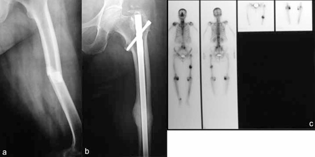
Figure 1. a) Antero-posterior radiograph of the left femur demonstrates a complete displaced transverse midshaft fracture with a small medial spike, b) 15 months after intramedulary nailing antero-posterior radiograph shows union and a significant amount of callus formation, c) bone scintigraphy shows the expected signs of bone healing.
Ten months post-operatively, the patient was referred to our centre. BMD measurements at the lumbar spine (LS: 0.800 g/cm-2, T-score= -3.2) and the right femoral neck (FN: 0.645 g/cm-2, T-score= -2.8) were compatible with osteoporosis. Biochemical evaluation revealed acceptable levels of 25(OH)vitamin D (28.2 ng/mL, reference value (r.v.) 20-58 ng/ml), while bone markers were within normal range (PINP: 26.8 ng/ml, r.v. 16.3-73.9 ng/ml; sCTX: 0.274 ng/ml, r.v. <0.73 ng/ml).
Eleven months following the fracture, given the history of AFF and significant residual fracture risk, TRP treatment was initiated along with continuation of calcium and vitamin D. Four months later, radiographic evaluation revealed complete fracture healing (Figure 1b, 1c). At 6 months of TRP treatment, increase in PINP levels confirmed adequate response (69.86 ng/ml, increase 160%). Eleven months after initiation of TRP she complained of pain to the contralateral (right) femoral diaphysis. The pain had been progressively worsening, becoming of the same intensity as the previous pain experienced before fracture (10/10). New bone scintigraphy revealed additional increased focal uptake in the right femoral mid-diaphysis (Figure 2a). The patient was advised to avoid weight-bearing by using crutches, while treatment with acetaminophen led to partial pain remission.
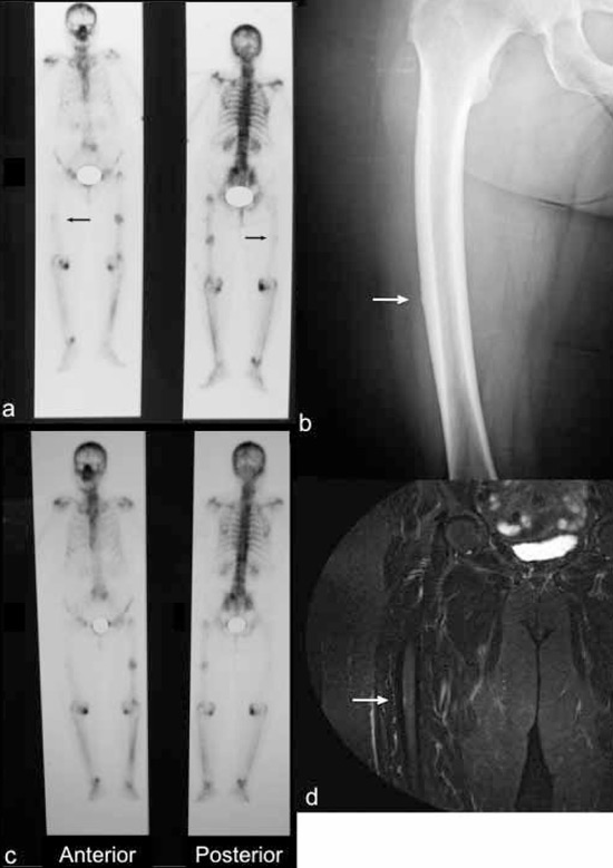
Figure 2. a) Bone scintigraphy 22 months after fracture showing increased uptake of radioisotope in the right femoral middiaphysis at the same level as the fracture site of the left femur (black arrow), b) antero-posterior radiograph of the right femur reveals subtle localized periosteal reaction of the lateral femoral midshaft (white arrow), c) 2 years after teriparatide treatment bone scan reveals partial reduction in radioisotope uptake in the right femoral midshaft and d) STIR MRI imaging revealed subtle linear intracortical hyperintensity at the right femoral diaphysis (white arrow).
At the completion of 2 years of TRP, the patient felt slight discomfort in the right femoral diaphysis (1/10), while at the left femur the pain had completely resolved. Radiograph of the right femur showed subtle localized periosteal reaction of the lateral femoral midshaft (Figure 2b). Scintigraphy showed a slight diminution of the periosteal reaction at the right femoral diaphysis (Figure 2c). Short TI Inversion Recovery (STIR) MRI imaging revealed subtle linear intracortical hyperintensity at the right femoral diaphysis (Figure 2d). BMD testing revealed increase at the LS 7.61% and 0.8% at the right hip.
Following the completion of the TRP course, 1-year SR treatment resulted in 9.2% further BMD increase at the LS and 1.4% at the right hip. She does not report pain or discomfort at the femurs (0/10) and she walks without limping. Radiographs and bone scintigraphy revealed almost the same findings, MRI imaging showed no periosteal and medullary signal abnormalities in the right mid-femoral shaft, while bone markers remain within normal range.
DISCUSSION
We report a patient who had a complete AFF and a contralateral incomplete femoral fracture after long-term BP administration, treated sequentially with TRP and SR with a favourable response. Both fractures, complete and incomplete, fulfilled all the major criteria of AFF as: 1) they were located at the femoral diaphysis and 2) were without history of trauma, while the complete fracture, 3) was non-comminuted, 4) with a transverse configuration and 5) extended through both cortices with a medial spike, whilst the incomplete fracture involved only the lateral cortex. Several minor criteria were also fulfilled as there was: 1) localized periosteal reaction of the lateral cortex and there were, 2) prodromal and 3) bilateral symptoms. Although our case fulfilled all the major criteria of AFF and several minor criteria, there are some distinguishing features, such as the emergence of the incomplete fracture even during TRP treatment, probably underlying the sustained effects of BPs, leading to bone fragility in the susceptible patient.
The clinical characteristics of atypical fractures, the occurrence in BP-naive patients and the location at sites other than the femur point to a systemic disorder with a different underlying pathogenesis from osteoporosis, probably resembling that of stress fractures.2 Also, biochemical bone turnover markers are often within the normal range. These similarities have set the stage for the application of full-length intramedullary nails that accomplish endochondral fracture repair, interruption of BP therapy, administration of calcium and vitamin D and institution of anabolic therapy, particularly TRP in the management of patients with AFF.3
Management of AFF with intramedullary nails is associated with a high failure rate reaching 50%,3 compared with typical femoral fractures, where the same procedure results in a 98-99% healing rate.7 Thus, close observation of the healing process is warranted, possibly with early institution of anabolic therapy.
The role of vitamin D deficiency in the pathogenesis of stress fractures and AFF has been emphasised, although the true prevalence is underreported. Burgi et al reported that 25(OH)D levels over 40 ng/ml were associated with half the risk of stress fractures at the tibia and fibula in female US navy recruits, while calcium and vitamin D administration has been shown to prevent stress fractures in the same population.8,9 In addition, although fracture healing is certainly delayed in cases of vitamin D deficiency, there is a paucity of studies directly addressing the effect of vitamin D on fracture healing. In any case, given that even vitamin D levels lower than 30 ng/ml might be related to mineralization defects,9 prompt substitution therapy is imperative for a favourable outcome.
The action of TRP includes stimulation of both modeling and remodeling-based bone formation followed by increase in bone resorption. Its effect on stress fracture healing was recently proved positive in a rat model of ulna stress fractures,10 while there have been only a few patients reported with AFF treated with TRP with a favourable outcome.4,5,11 Several studies indicate that TRP reduces microdamage accumulation and decreases matrix mineralization and collagen cross-link ratio, thereby attenuating the alleged adverse effects associated with prolonged BP administration.12 Finally, recent evidence indicates that TRP exerts bone anabolism, via a VEFG-dependent mechanism, by localizing capillaries near sites of new bone formation.13 Thus, there is good evidence that TRP would probably promote AFF healing and reduce the risk of such fractures in susceptible patients. In our case, despite previous BP treatment, TRP resulted in a favourable response in terms of both bone markers and BMD, and healing of contralateral incomplete AFF.
SR is characterized by a unique mode of action, decreasing bone resorption and concurrently increasing or at least maintaining bone formation.14 It reduces the risk of vertebral and non-vertebral fractures in patients with postmenopausal osteoporosis14 and improves trabecular and cortical structures15 as well as intrinsic bone properties,16 while having neutral effects on mineralization.17 Recent evidence indicates that under conditions of impaired fracture healing, such as ovariectomy induced bone loss, SR exerts beneficial effects on the healing process, including increasing bone strength at the fracture site.18 Although there are no animal studies concerning the effect of SR on stress fracture healing, a recent study reported a favourable outcome in two patients with AFF treated with SR.5 Moreover, SR has some additional advantages such as post-hoc based evidence of hip fracture risk reduction in patients with high risk.19 Thus, in patients with AFF treated in the past with TRP, having other contraindications to TRP, or even displaying significant future risk for hip fracture, SR treatment seems the most rational option. Finally, although there is clear evidence that anticatabolic therapy following a TRP course preserves or further improves BMD, in the setting of AFF, restarting BP treatment might result in re-emergence of atypical skeletal fragility. Accordingly, SR is probably the only available treatment option, especially in cases with significant ongoing fracture risk. At least in our case, SR resulted in a favourable response in terms of BMD, while bone markers remained within normal limits.
In conclusion, our case indicates that sequential therapy with TRP and SR in cases of AFF might be a rational treatment option. However, in the absence of controlled trials, if ever conducted, we cannot be certain whether the fractures healed spontaneously with the general recommended management in such cases. Therefore, there is a need for additional information concerning the effect of TRP and SR, given alone or sequentially, in patients with AFF in order to incorporate these drugs into the management plan of AFF.
ACKNOWLEDGMENTS
Dr. Kalliopi Lampropoulou-Adamidou and Dr. Symeon Tournis equally contributed to the design and preparation of the manuscript.
CONFLICT OF INTEREST
The authors declare that there is no conflict of interest that could be perceived as prejudicing the impartiality of the review reported.
FUNDING
This research did not receive any specific grant from any funding agency in the public, commercial or not-for-profit sector.
REFERENCES
1. Papapetrou PD, 2009 Bisphosphonate-associated adverse events. Hormones (Athens) 8: 96-110.
2. Compston J, 2011 Pathophysiology of atypical femoral fractures and osteonecrosis of the jaw. Osteoporos Int 22: 2951-2961.
3. Shane E, Burr D, Ebeling PR, et al, 2010 Atypical subtrochanteric and diaphyseal femoral fractures: report of a task force of the American Society for Bone and Mineral Research. J Bone Miner Res 25: 2267-2294.
4. Gomberg SJ, Wustrack RL, Napoli N, Arnaud CD, Black DM, 2011 Teriparatide, vitamin D, and calcium healed bilateral subtrochanteric stress fractures in a postmenopausal woman with a 13-year history of continuous alendronate therapy. J Clin Endocrinol Metab 96: 1627-1632.
5. Carvalho NN, Voss LA, Almeida MO, Salgado CL, Bandeira F, 2011 Atypical femoral fractures during prolonged use of bisphosphonates: short-term responses to strontium ranelate and teriparatide. J Clin Endocrinol Metab 96: 2675-2680.
6. Anastasilakis AD, Goulis DG, Polyzos SA, et al, 2009 No difference between strontium ranelate (SR) and calcium/vitamin D on bone turnover markers in women with established osteoporosis previously treated with teriparatide: a randomized controlled trial. Clin Endocrinol (Oxf.) 70: 522-526.
7. Weil YA, Rivkin G, Safran O, Liebergall M, Foldes AJ, 2011 The outcome of surgically treated femur fractures associated with long-term bisphosphonate use. J Trauma 71: 186-190.
8. Lappe J, Cullen D, Haynatzki G, Recker R, Ahlf R, Thompson K, 2008 Calcium and vitamin d supplementation decreases incidence of stress fractures in female navy recruits. J Bone Miner Res 23: 741-749.
9. Priemel M, von Domarus C, Klatte TO, et al, 2010 Bone mineralization defects and vitamin D deficiency: histomorphometric analysis of iliac crest bone biopsies and circulating 25-hydroxyvitamin D in 675 patients. J Bone Miner Res 25: 305-312.
10. Sloan AV, Martin JR, Li S, Li J, 2010 Parathyroid hormone and bisphosphonate have opposite effects on stress fracture repair. Bone 47: 235-240.
11. Chiang CY, Zebaze RM, Ghasem-Zadeh A, Iuliano-Burns S, Hardidge A, Seeman E, 2013 Teriparatide improves bone quality and healing of atypical femoral fractures associated with bisphosphonate therapy. Bone 52: 360-365.
12. Paschalis EP, Glass EV, Donley DW, Eriksen EF, 2005 Bone mineral and collagen quality in iliac crest biopsies of patients given teriparatide: new results from the fracture prevention trial. J Clin Endocrinol Metab 90: 4644-4649.
13. Towler DA, 2011 Skeletal anabolism, PTH, and the bone-vascular axis. J Bone Miner Res 26: 2579-2582.
14. Marie PJ, Felsenberg D, Brandi ML, 2011 How strontium ranelate, via opposite effects on bone resorption and formation, prevents osteoporosis. Osteoporos Int 22: 1659-1667.
15. Arlot ME, Jiang Y, Genant HK, et al, 2008 Histomorphometric and microCT analysis of bone biopsies from postmenopausal osteoporotic women treated with strontium ranelate. J Bone Miner Res 23: 215-222.
16. Ammann P, Badoud I, Barraud S, Dayer R, Rizzoli R, 2007 Strontium ranelate treatment improves trabecular and cortical intrinsic bone tissue quality, a determinant of bone strength. J Bone Miner Res 22: 1419-1425.
17. Farlay D, Boivin G, Panczer G, Lalande A, Meunier PJ, 2005 Long-term strontium ranelate administration in monkeys preserves characteristics of bone mineral crystals and degree of mineralization of bone. J Bone Miner Res 20: 1569-1578.
18. Habermann B, Kafchitsas K, Olender G, Augat P, Kurth A, 2010 Strontium ranelate enhances callus strength more than PTH 1-34 in an osteoporotic rat model of fracture healing. Calcif Tissue Int 86: 82-89.
19. Reginster JY, Seeman E, De Vernejoul MC, et al, 2005 Strontium ranelate reduces the risk of nonvertebral fractures in postmenopausal women with osteoporosis: Treatment of Peripheral Osteoporosis (TROPOS) study. J Clin Endocrinol Metab 90: 2816-2822.
Address for correspondence:
Symeon Tournis, M.D., PhD., Laboratory for the Research
of Musculoskeletal System “Th. Garofalidis”, Medical School, University of Athens, General Hospital of Athens KAT, Greece, 10 Athinas Str., Kifissia, PC: 14561, Athens, Greece, Tel.: +30 2108018123, Fax: +30 2108018122,
E-mail: stournis@med.uoa.gr
Received 17-09-2012, Accepted 30-01-2013