1Department of Cytopathology, Naval Hospital of Athens, Athens, 2Department of History of Medicine, Ioannina University School of Medicine, Ioannina, Greece
In 1847 Felix-Archimede Pouchet effectively launched the study of the physiology of cytology. Now 160 years later, the authors briefly trace the development of hormonal cytology to our present knowledge and practice. In the course of the paper the contribution of George Papanicolaou is stressed because of his monumental contribution to a major segment of medical practice of great emotive import.
Cells, Ηormonal cytology, Papanicolaou, Pouchet, Stockard, Vaginal smears
INTRODUCTION
The study of the cytology of vaginal smears was initiated in the middle of the 19th century by Felix-Archimede Pouchet in Paris (1847), who studied the morphological changes associated with the regular oestrous cycle. He described the microscopic variations in vaginal secretions during the different phases of the menstrual cycle and in the period between two consecutive menstruations.1 Although Pouchet was not able to distinguish the fine differences between the intermediate and the well differentiated superficial (karyopycnotic) cells, he took the initial step in the field of functional (hormonal) cytology.
THE TWO PERIODS
The history of Hormonal Cytology could be divided roughly into two periods.
The first period commenced in the middle of the 19th century and extended to the middle of the 20th century. During this timeframe certain observations were made, such as Pouchet’s, which marked its inception.
George Papanicolaou launched the first important hormonal cytology development. His monumental contribution was applied to many practical uses and he was unreservedly acknowledged as the founder of a new medical specialty, diagnostic cytology and, by extension, Hormonal Cytology.2
In 1917 George Papanicolaou was working as an assistant in the Anatomy Section of the Cornell Medical School, under the direction of Professor Charles Stockard. Papanicolaou resolved to continue and complete his experimental research on the determination of sex that he had embarked upon while studying in Germany. Papanicolaou studied guinea pig vaginal smears with particularly encouraging results.
In the middle of the second decade of the 20th century, Stockard and Papanicolaou3 first used microscopic observation of vaginal smears in the study of the oestrous cycle of animals. One decade later, Ramirez (1928) extended this research effort to humans, studying cycle variations caused by changes in ovarian hormones.4
Two relevant papers published by Papanicolaou and Stockard (1917), one in Science entitled “A rhythmical ‘heat period’ in the guinea pig”5 and the other in the American Journal of Anatomy entitled “The existence of a typical oestrous cycle in the guinea pig: with a study of its histological and physiological changes”3, were to be the springboard for all further developments in the field of Hormonal Cytology.
The original and pioneering studies of the great researcher towards the determination of the hormonal cycle of mammals were to be concluded with the study of cytomorphology of vaginal smears in direct relation to the hormonal changes of the organism. George Corner, a famous researcher in the field of reproductive endocrinology, indicated that the discovery of the ovarian hormones by Edgar Allen and Edward A. Doisy* in 1923 was based on the method first described by Papanicolaou. According to Corner “the connection of the knowledge of the cycle of the pig and the reproduction of the rabbit led indirectly to the discovery of the hormone of the corpus luteum and furthermore to a more complete and clearer understanding of the whole phenomenon than in the past”.6
* Edward Doisy and Henrik Dam received the 1943 Nobel Prize for Physiology/Medicine for their work on Vitamin K.
Papanicolaou later invented his own procedure of smearing and permanence as a means of describing the maturing stages of the epithelial cells. Among his numerous observations on distinct cell morphology, he noted the navicular shape of the cells during pregnancy, which he named “boat-shaped cells”7.
The publication of Papanicolaou’s paper in the American Journal of Anatomy in 1933 entitled “The sexual cycle in the human female as revealed by vaginal smears”8 denotes the first period of the history of Hormonal Cytology during which Papanicolaou established the study of vaginal smears as a hormonal test to describe the genital cycle of the female. In this paper Papanicolaou described the cell morphology, the staining method, which today bears his name and is used in all cytology laboratories worldwide, and divided the monthly cycle into four phases: the menstruation phase which extends from the 1st to the 7th day, the connecting phase from the 8th to the 12th day, the follicular phase from the 13th to the 17th day and the premenstruation phase from the 18th day until the start of the period, as well as an ovulation stage between the 12th and the 13th day of the cycle.
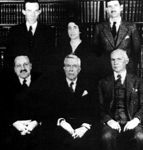
Figure. Teaching staff of the Anatomy Department of Cornell Medical School. First row from left: Dr. George N. Papanicolaou and Prof. Charles R. Stockard.
At about the same period, studies of other researchers like Dierks (1927) and Murray (1938) confirmed the views of Papanicolaou histologically and cytologically, respectively3.
At the beginning of the ’40s, E. Shorr9 modified the Papanicolaou staining method, aiming at a fast evaluation of the hormonal status, and towards the end of the decade two other pertinent papers were published. First the illustrated publication “La citologia vaginal humana” (Human vaginal cytology) by Argentineans Allende and Orias (1947) in Spanish, later (1950) translated into English.4 Following this came the publication of the paper by Papanicolaou, Traut and Marchetti (1948) “The epithelia of woman’s reproductive organs”4. The last two papers served as the impetus for the global implementation of the cytological method as a painless, quick, economical and bloodless approach for the evaluation of the ovarian function.
The second period covers the years from the beginning of the ’50s to our time. Throughout the years before and after second World War, for a period of more than 20 years, the cytological examination of vaginal smears was the main method used for the hormonal testing of women’s cycle. The method developed by Papanicolaou attracted many followers and was implemented extensively.
J. Pundel (1952) defined the criteria of the oestrogenic, luteinizing and androgenic influence on the vaginal epithelium, and defined with precision the day of ovulation. He also distinguished the anovulation cycles as hypo- and hyper-follicular, using the “eosinophilic index”, which expresses the percentage of cells with eosinophilic cytoplasm. Pundel described seven phases in the monthly cycle which relate to the histological image of the endometrium.10
O. Nyclicek (1951) introduced a new system which included a “maturity index” and which remains till today the most popular method in cytology for evaluation of hormonal status.11
G. Pincus and his associates (1955) and later R.F. Brown and his associates (1959) investigated urinary estrogen levels, confirming the declining values of hormones in perimenopausal women.12
The International Academy of Cytology (1958) published a terminology of cells and indices to be used for classification of hormonal status. The superficial cell indices, karyopycnotic and eosinophilic, remained very popular for many years.12
In the following years the work of other researchers questioned the diagnostic accuracy of the vaginal smears in the study of the menstrual cycle. Never¬theless, the study of vaginal smears worked as a supplement to the existing biochemical methods (such as the quantitative determination of steroids in urine or blood) dealing with the etiology of menstrual irregularities. Prior to the introduction of this method, biopsy of the endometrium was the only diagnostic approach in cases of amenorrhea.
Thus, from the end of the ’50s the examination of vaginal smears was gradually abandoned for the evaluation of the monthly cycle, while it continues to be used for the control of administration of hormonal preparations as well as for the early identification of cancer of the uterine cervix.
REFERENCES
1. Grunze H, Spriggs A 1980 Founders of Clinical Cytology. In: Grunze H, Spriggs A (eds) History of Clinical Cytology, G-I-T Verlag Ernst Giebeler; pp, 39-40.
2. Louros NC, Marketos SG, 1983 George N. Papanicolaou (1883- 1962). Discoverer of the “Pap Test”. World Health April-May: 23-25.
3. Stockard CR, Papanicolaou GN, 1917 The existence of a typical oestrous cycle in the guinea pig: with a study of its histological and physiological changes. Amer J Anat 22: 225-283.
4. Grunze H, Spriggs A 1980 Cytology in Gynaecology (“Hormonal Cytology”). In: Grunze H, Spriggs A (eds) History of Clinical Cytology, G-I-T Verlag Ernst Giebeler; pp, 91-92.
5. Stockard CR, Papanicolaou GN, 1917 A rhythmical “heat period” in the guinea pig. Science 46: 42-44.
6. Carmichael ED, 1984 Dr. Papanicolaou and the Pap smear. Ala J Med Sci 21: 101-104.
7. Papanicolaou GN, 1925 The diagnosis of early human pregnancy by the vaginal smear method. Proc Soc Exp Biol Med 22: 436-437.
8. Papanicolaou GN, 1933 The sexual cycle in the human female as revealed by vaginal smears. Amer J Anat 52: 519-637.
9. Shorr E, 1940 A new technique for staining vaginal smears II. Science 91: 579-580.
10. Pundel JP, 1959 Vaginal cytology at the end of pregnancy. Acta Cytol 3: 253-263.
11. Nuclicek O, 1951 Importance of vaginal cytogram for diagnosis and therapy in the deficiency of oestrogenic hormones. Gynaicologia 131: 173-183.
12. O’ Dowd MJ, Philipp EE 1994 The menopause. In: O’ Dowd MJ, Philipp EE (eds) History of Obstetrics and Gynaicology, Informa Health Care; pp, 318.
Address for correspondence:
Diamantis Aristidis, 18, Agamemnonos Str., 15561, Cholargos,
Athens, Greece, Tel.: +30 210 7261154,
Fax: +30 210 7261397, e-mail: aristidis.diamantis@gmail.com
Received 08-06-07, Revised 08-09-07, Accepted 20-10-07